Such good advice from my creative mentor Samantha Bennett of therealsambennett.com . Start with who you know. Start with something you’re passionate about and good at doing. Start with something that gives value or solves a problem for the people you want to help the most. Make something no matter if it’s a “B”, then get it out to your chosen market so you can get feedback and improve it.
Here’s how I’m getting on. In this post I’ll share what gear I’m using to take my images. Take a look at the photo to see what I mean.
But going back a step. You might want to know the reason why I’ve chosen this particular project. Over the years, I’ve spent many hours poring down a microscope searching and counting fungal spores. Specifically the hand grenade shaped ones that are characteristic of a fungus called Pithomyces Chartarum. Following the trend of spore counts in a set of sampling paddocks or fields on a dairy farm can help with making decisions on when to take action.
Facial Eczema is a nasty condition. You might think that it’s a skin disease. Yes, in many cases a dairy cow with this disease will have obvious signs of photosensitization with white patches on her coat going red and eventually peeling off. But Sporidesmin, which is the toxin the spore produces, actually does more damage to a cows liver (or sheep or goat). It’s this liver pathology that can kill her or make her very sick.
Counting spores is a relatively simple process. But it does get tedious. This is why I want to automate it in some way. I’d like to make it faster and easier to do so more dairy farmers will monitor more of their paddocks. This would give them the most up to date information on their own farm in their own micro climate. I’ll start with an algorithm that can correctly identify a P.Chartarum spore from something that isn’t. This is a start. It’s one piece of the puzzle.
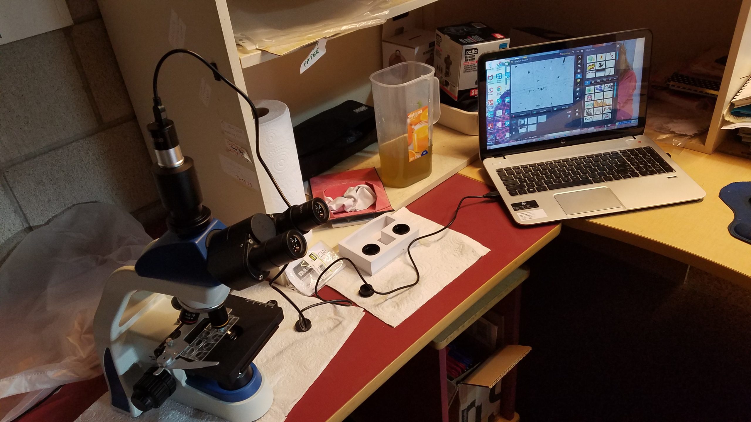
Perhaps my journey will inspire you to start your own image classification work. What do you need? First, a Trinocular Biological Microscope. Mine is an E3 series one I bought from Magnifiers in New Zealand. Click this link to find out more about it.
You’ll also need a camera of some sort to take the images with. I bought a Video Camera from the same company. A really really important bit to make sure you have is an adapter that fits between the trinocular head and the piece that takes the pictures. Don’t be like me and spend hours trying to figure out how the camera fits in the trinocular head! The adapter should come with your microscope camera. Then all you need to do is connect the camera to your laptop via the USB lead and hey presto!
The last step is to change the settings in your laptop camera software program. Mine is CyberLink YouCam. You need to do this so it captures the photos you take of spores under the microscope and not the images of you squinting at your computer screen. Click the settings button to the right of the screen. Your microscope camera should show up in the “Capture Device” drop down menu. Choose it and save the settings. If you need to check on what it is called go into Device Manager and find the camera in “Imaging Devices”.
You will probably have a different version of CyberLink YouCam. My laptop is an old one, like me, and I run my HP Envy with Windows 8.
So far I’ve taken 92 photos with this set up and I’ve created a simple Xcel Spreadsheet called “labels”. I’ll convert it into a csv file later. There are two columns. One is named “id” and has the image file name for identification e.g. Snapshot_20220517. The other column I’ve headed up as “classification”. Photos of P. Chartarum spores I classify as “1”or Yes. And images of other things I see in the pasture wash grid I choose “0” or No. I want to have roughly the same number of “1”s and “0”s in the final dataset of 1000.
Only 908 more photos to take!